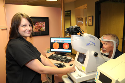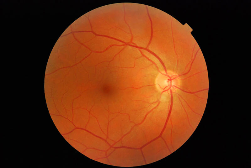
The retina camera takes a photograph of the retina. The retina is the tissue that lines the inside of the eye and contains the rod and cone photoreceptor cells.
When looking at the picture of the retina, the yellow circle to the right is the optic nerve. It is where all of the nerves come together to leave the eyeball. The blood vessels also travel through the optic nerve. The artery is the skinnier red vessel that brings the blood into the eye. And, the vein is the thicker red vessel that drains the blood out of the eye.
 The macula is the darker area in the center of the photo. It is the part of the eye that is responsible for your clear central vision.
Many different conditions that affect the body can also affect the retina, such as diabetes, and hypertension. If these conditions are affecting your eye, you will be able to see them in the retina photograph. Other common eye conditions, such as macular degeneration and glaucoma, can be seen in the retina photograph.
Our retina camera is the new Topcon NW8, an auto-focus, auto-shoot digital camera that takes amazingly clear, detailed retina photographs in just seconds. For many patients, the photo can be taken through an un-dilated pupil.
Dr. Davidson and Dr. Keltner highly recommends that the retinal photographs be taken to document a healthy baseline or to help monitor for changes. It also gives her a full view of the back of the eye so that she can study it and explain the photograph to you in great detail.
Unfortunately, most insurance companies do not yet cover retina photography for healthy eyes.
Request an Appointment
Schedule your appointment today with Riverside Optometry or call us at (951) 784-2420. We are excited to serve you and your family!


